Immediately after the birth of a child, you can notice such spots on the skin that show maximum activity in growth in the first six months. Hemangioma occurs in 10% of newborns, several times more often - in girls. After the 1st year of life, tumor growth slows down and its involution occurs - a gradual disappearance. By the age of five, 50% of hemangiomas disappear, and by the age of seven - up to 70% (early involution). Since hemangioma is a hormone-sensitive tumor, its complete inhibition occurs at puberty (late involution).
Why does hemangioma occur?
The reasons
The true causes of these tumors are not yet known. It is assumed that triggering factors in their development may include:
- ARVI in the mother during pregnancy for a period of 3 to 6 weeks, when the cardiovascular system forms in the embryo;
- Rhesus - a conflict in the mother during pregnancy;
- taking medication, alcohol, or smoking while carrying;
- hormonal disorders in a pregnant woman or in a child;
- poor environmental conditions;
- burdened by heredity.
Types of hemangiomas
Depending on the structure and level of location, the following types of hemangiomas are distinguished:
Cavernous hemangioma
This type of tumor also occurs in organs with abundant blood supply: liver, spleen, kidneys, adrenal glands, lungs and brain.
Cavernous hemangioma in a newborn in the liver is considered very dangerous. In most cases, it makes itself felt only with the appearance of complications or accidental diagnosis, since it is asymptomatic in the body. In case of trauma, hemangioma may rupture, and hemorrhage arising under this under the capsule of the liver or into the abdominal cavity, in 80% or more cases, leads to death.
The spleen is a very well vascularized organ, therefore, hemorrhages in it due to rupture of hemangiomas are most dangerous, since they are profuse in nature.
Brain cavernous hemangioma is one of the most insidious tumors. Despite its good quality, tearing it leads to intracerebral or subarachnoid hemorrhage, which entails a deep coma or death.
Capillary hemangioma
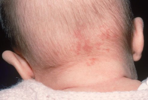
Capillary (simple) hemangioma is formed from the vessels of the dermis and never affects the underlying layers of the skin (with the exception of the combined type of hemangioma). The structure is capillary vessels intertwined into a ball. The tumor protrudes slightly above the surface of the skin, extremely rarely gives hemorrhages. As a rule, it is small in size, about 1 cm in diameter. A tumor is considered the more favorable, the paler its color - this indicates its involutional development.
Combined hemangioma
The combined hemangioma is represented by simple and cavernous parts and is insidious in that it is often mistaken for a capillary tumor, while its cavernous part can be fraught with danger.
A mixed tumor occurs when, along with a vascular neoplasm, tumor cells of the connective, nervous or other tissues are present.
Video
Diagnosis and treatment of hemangiomas
The choice of treatment measures depends on the results of diagnostic studies of hemangiomas. First of all, the doctor must differentiate the tumor from other diseases. It can be squamous cell carcinoma or glomus angioma. Hemangioma is sometimes similar to vascular malformations, some forms of nevi and cysts, pyogenic granuloma.
After diagnosis, the decision on subsequent treatment is made depending on the progression of the tumor.
In the period up to 1 month (neonatal period) surgery is excluded. Monitoring of the "behavior" of hemangiomas. If the tumor does not change in size and color, then before visiting a kindergarten, it is simply observed by a specialist. If it increases, disrupts the functioning of organs or poses a threat to life, then measures are taken to remove it. As a rule, such operations are prescribed for a child at the age of 3 months, six months or 1 year.
Hemangiomas are treated in the following ways:
- Surgery includes complete or partial excision of the tumor and is indicated for its rapid progression, provided that the operation does not cause a severe cosmetic defect and does not interfere with the functioning of the organs. Surgical treatment is preferred in case of large tumor sizes, but then after resection it becomes necessary to take a donor skin flap from another part of the body. This is especially true when treating hemangiomas on the scalp or eyelid. In children, such operations are performed only in exceptional cases with parallel blood transfusion, under general or local anesthesia.
- Conservative treatment:
- Cryotherapy (snow from carbon dioxide): applied to small-sized hemangiomas (2 -2.5 cm). Snow is applied to the tumor site with the capture of healthy tissue up to 0.5 cm. After this, a depressed surface is formed, which soon swells, turning into a bubble. Then a crust forms, which disappears after two weeks.
- Injection treatment with a sclerosing effect on the vessels of the tumor, after which connective tissue is formed in its place. For injection, 70% alcohol and a solution of quinine - urethane are used. With a few injections, an infiltration roller forms first around the tumor, then in its center. Once a week, repeat the procedure after the swelling disappears. This method is used when it is impossible to conduct surgical treatment, due to the difficult location of the tumor: eyelid, oral mucosa.
- Electrocoagulation it is used to treat small (not more than 5 mm) capillary, cavernous and stellate hemangiomas, as well as to remove the remaining parts of the tumor after other procedures. Under the influence of electric current, the tumor tissue coagulates, after which a crust forms, which independently passes with time.
- Radiation therapy used in the treatment of subcutaneous cavernous hemangiomas or tumors that are localized on internal organs. Radiotherapy has a negative effect on the whole body, so it is not used for hemangiomas in newborns. Radiation therapy is prescribed exclusively after 6 months.
Video
Hemangioma Complications
In addition to the fact that hemangiomas can cause hemorrhages or disrupt organ function, they also contribute to the formation of blood clots in the tumor cavity. Because of this, the consumption of platelets in the body increases, which leads to a deterioration in blood coagulation. This is characterized by the Kazabakh – Merrit symptom complex — a disease of newborns and children up to a year old who have large hemangioma.
If the tumor is located in traumatic areas or in the genital area, it is often subjected to ulceration.
Hemangioma in newborns is most often limited to a cosmetic defect or completely disappears. But, nevertheless, the presence of a tumor in a child obliges parents to provide the baby with systematic observation by a specialist.


Hemangioma is now very common among children. This also did not pass us by. At my daughter right after birth I noticed a red spot on my forehead. The doctor said that in most children this passes without a trace with age. Of course, I worried a lot about this. But, literally, a year later she completely disappeared. So it’s not so scary!
My daughter is already 17 years old, and the red spot on her forehead has still not passed. So who does it like ...
You have a stain, and my niece has a bump near the elbow, quite large for its size.
Good day!
The daughter was born without a single spot, after about two
weeks, a small red dot appeared on the head in the soft spot area, which by the month and a half grew up to almost a centimeter in circumference, became very bright and bumpy (photo 1).
We were very worried, showed the daughter to many doctors, everyone made a diagnosis - hemangioma. Who said to cut, who did not touch.
A very good surgeon advised treatment with aniprilin.
My wife and I decided not to rush and wait at our own risk.
We are now 1.3 years old, hemangioma has almost disappeared (photo 2).
Decide for yourself how to be!
Hemangioma appeared after the maternity hospital on the cheek, at 2 months it began to grow actively. In 4 months went to the regional clinical hospital in g.Dnepropetrovsk for 3 days, took propronalol 3 times a day. Then at home anaprilin (calculated by weight) and once a month we go to the control. Now the child is 8 months old. - still on tablets, the tumor is not visible at all, barely pink spots are noticeable. Satisfied.
In rare cases, hemangiomas can affect the arteries, forming in the form of large formations with many cavities that impede the normal functioning of the circulatory system.
Hello, my baby got a hemangioma. After discharge, approximately 2 weeks. We are now five months old. Cauterized for two months. 4 months did the injection. Hemangioma as it was and still is. Swollen ball. Now I do not know what to do? Hemangioma is right on the neck. Doctors are afraid to operate. Close to the carotid artery. Help, what should I do please?
Hello! Help who came across!
The baby was born immediately covered with a haemongioma. The whole body, except the head. What to do?
We have hemangioma on our neck, we don’t have surgery for 6 months, it’s dangerous, it has grown 4 months to a decent size, we don’t know what to do from Bishkek ourselves, we don’t have such a clinic, can someone tell me who came across, a good clinic .